
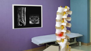
What is Spinal Stenosis?
The spine consists of bones called vertebrae. The job of these bones is to provide stability and support for the upper body. Nerves run through the openings in the vertebrae and transmit signals from the brain to the rest of the body. If the surrounding bones or tissues get damaged, it can affect basic movements like walking, balance, and sensation.
Spinal stenosis is a condition that causes the spinal column to narrow and compresses the spinal cord. This does not happen quickly, but is a more gradual concern. Too much narrowing compresses the nerves in the spinal and causes problems.
While aging is the most common cause of spinal stenosis, conditions like osteoarthritis and rheumatoid arthritis can contribute to the pressure on your spinal cord.
How is Spinal Stenosis Diagnosed?
What are the Symptoms?
Symptoms can vary depending on the location of the stenosis and the nerves that are affected. Since spinal stenosis is a gradual onset condition, the symptoms tend to start slowly and worsen over time. In fact, spinal stenosis may appear on an MRI or a CT scan without the patient having any symptoms at all.
Cervical Spine
- Numbness or tingling in the hand, arm, foot, or leg
- Difficulty walking or with balance
- Neck pain
- Bowel or bladder conditions such as urinary urgency or incontinence
Lumbar Spine
- Weakness in the foot or leg
- Pain or cramping in one or both legs after long periods of standing or walking
- Back pain
- Numbness or tingling in the leg or foot
How is Spinal Stenosis Diagnosed?
Your doctor will diagnose spinal stenosis by reviewing your medical history and perform a physical examination. They will also want to discuss any symptoms you experience. After a full review, your doctor may order several imaging tests to pinpoint the cause of symptoms and signs.
Imaging tests may include:
X-Rays
X-rays can determine if there was a bone change that could affect the spinal column, such as bone spurs.
Magnetic Resonance Imaging (MRI)
MRIs utilize a powerful magnet and cross-sectional radio waves to produce images of your spine. MRIs detect issues with ligaments and discs. It also has the ability to determine if there is a tumor pressing on the nerves. The most important reason why MRIs are helpful is that they are able to show where the nerves in the spinal cord are being compressed.
CT Scan
If you are unable to have an MRI, a CT scan may be used instead. This type of imaging test combines X-Ray images that are taken at many different angles to create a detailed, cross-sectional image of your body.
Get started on getting the relief you deserve by fill out the form below. The team at Progressive Pain Management is here to help you reduce pain and enhance your quality of life.